Pre-Stained Morphology SlidesDRM-900
These slides are used for the staining of spermatozoa, blood, CSF, and urine cytology. Each slide contains a dried layer of stain that once in contact with cells, facilitates morphology assessment. Cell-VU® provides cleaner and clearer slides for rapid cellular visualization and differential cell assessment.
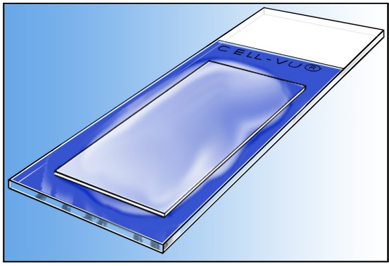
CELL-VU® Pre-stained Morphology slides combine conventional microscope slides with a proprietary combination of methylene blue and cresyl violet stains into a single, ready-to-use device.
Samples are applied directly to the slide and a coverslip is placed over the sample. One step process limits exposure to reagents and potentially harmful stains.
CELL-VU® is used for World Health Organization and Kruger strict morphology sperm assessment from undiluted semen.
![]() Available for sales in the European Union
Available for sales in the European Union


Directions for Use - PreStained Morphology Slides
STEP 1
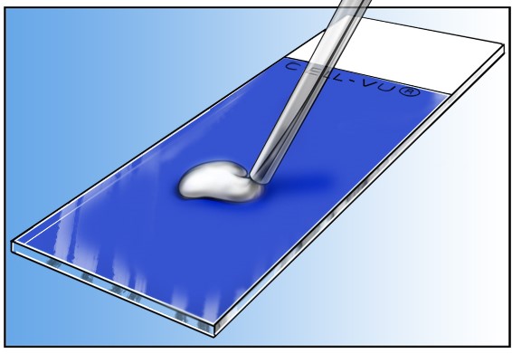
Apply a small drop (approximately 2-4 microliters) of specimen on the center of the slide.
NOTE: EDTA is the recommended anticoagulant for venous blood. Flouride oxalate and heparinized blood are NOT suitable.
STEP 2
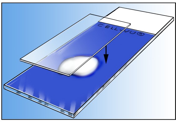
Place the coverglass (24 x 24 mm or 24 x 40 mm) on top of the slide, exerting slight pressure on the coverglass.
STEP 3
Allow the slide to stand at room temperature for 15 minutes before microscopic observation is made. The slides are used for evaluating sperm head/acrosome size, differentiation of white blood cells in peripheral blood and cytology of body fluids.
CELL-VU® Microscope Slides and Coverslips are used by trained laboratory professionals to assist with count and analysis of cells in body fluids, such as semen, blood, CSF and urine. The slides are used with a microscope in a laboratory setting.
Materials provided with DRM-900: Microscope Slide (725) 3x1 in with printing on top of the slide and a dried stain on the top of the slide.
Materials not provided but necessary: One standard coverslip per test.
Manufacturer: Millennium Sciences, Inc. 6899 Collins Ave, Unit 2908, Miami Beach, FL 33141 USA
CE Mark Authorized Representative: AR Experts BV, Boeingavenue 209, 1119 PD Schiphol-Rijk, The Netherlands, www.ar-experts.eu
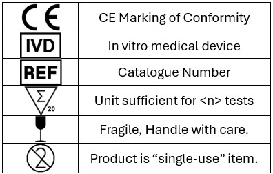
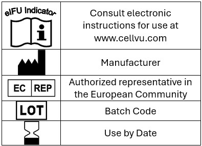
SYMBOL DEFINITIONS